Upper Arm
Proximal Humeral Fracture
Definition:
The proximal or shoulder-joint humeral fracture accounts for 5% of all fractures and is a typical fracture of the elderly. Often it is associated with osteoporotic bone changes, so that even a small trauma leads to breakage. It occurs in younger patients, usually as a result of a high speed impact injuires.
The most common form is the subcapital humeral fracture just below the humeral head, but also the humeral head can be directly affected.
Symptoms:
In addition to a painful restriction of movement, there is a pressure pain over the humeral head. In some cases, bruising occurs around the armpit, upper arm and lateral chest wall. Rarely, the axillary nerve is injured, which manifests itself as a disturbance of lateral shoulder sensation and loss of power in parts of the shoulder muscles.
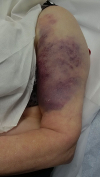
Picture: You can see a typical bruise on the area of the upper arm after a shoulder joint upper arm fracture..
Diagnosis and Therapy:
The first indication of a proximal humeral fracture is provided by the accident. Typical is a fall on the shoulder or the arm, also as indirect trauma with fall on the outstretched hand or the elbow. Furthermore, a physical examination is indispensable for the diagnosis of the above symptoms. The shoulder should be x-rayed in 3 planes. Sometimes, a supplementary computer tomography is necessary for further diagnosis and exact determination of the course of the fracture. Magnetic resonance imaging is only performed if there is a suspicion of a dislocation of the shoulder joint or a malignant underlying disease as the cause of the fracture.
There are many different classifications for the codification of proximal humeral fractures. A frequently used classification is NEER. Depending on the displacement, tilting and exact localization of the fracture clefts, the classification is divided into a total of 6 classes (NEER I - VI).
The classification of the fracture then determines the further procedure. If the fracture is submerged and not or only slightly displaced, conservative therapy can be applied. This usually applies to the frequent form of subcapital humeral fracture and thus to over 80% of fractures. The therapy includes wearing an immobilizing shoulder vest for 1 - 2 weeks. Long-term immobilization can lead to stiffening of the shoulder joint.
Surgical treatment should be performed for all initially or secondarily displaced fractures. Further indications for surgery include multi-piece fractures, open fractures, dislocation fractures or damage to nerves and vessels. Without surgical treatment, the shoulder may otherwise heal in a defective position or bone may not heal, which in turn can lead to severe functional and movement restrictions of the shoulder.
One possibility of surgical therapy is stabilization with a stable-angle plate In addition, with the frequent form of subcapital humeral fracture, a nail can be inserted into the upper arm and the fracture can thus be splinted from the inside. In case of severe damage, the humeral head may have to be replaced by a prosthesis.
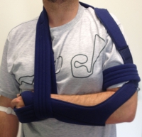
Picture: Shown is a model of a shoulder vest. The patient has suffered a fractured upper arm. Until the operation, the arm is immobilized by means of the shoulder vest.
After Treatment:
In conservative therapy, a timely start of physiotherapy after the first 1 - 2 weeks is important to avoid stiffening of the shoulder joint. Initially, starting with pendulum movements out of the immobilized shoulder vest, then training with increasingly active movement exercises. Regular positional examinations of the fracture by means of X-ray images are necessary in order to be able to detect any delay in time and, if necessary, to be able to treat it surgically.
After a successful operation, the follow-up treatment depends on what was implanted. In general, the physiotherapeutic exercise can begin early, in order to strengthen the muscles and restore mobility in the shoulder joint.
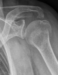
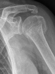
Picture: The subcapital humeral fracture shown here in the axis is not broken and only slightly bruised, so that a conservative therapy is possible. The left image shows the follow-up after 3 weeks. The patient has already started physiotherapy. The x-ray shows an increase bone healing.
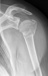
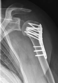
Picture: Here is a subcapital upper arm break in a young woman. An angle stable plate osteosynthesis was performed to stabilize the head in correct position to the humeral shaft and shoulder joint. Especially in young patients, We strive to treat non-invasively and as gently as possible. This possibility is often due to the bone quality in young as opposed to older people.

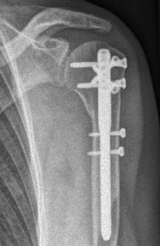
Picture: In the above pictures, a slightly shifted subcapital humeral fracture was internally splinted using intramedullary nail. The left picture is a follow-up examination half a year after the accident and shows the complete healing of the fracture with correct position of the bone.
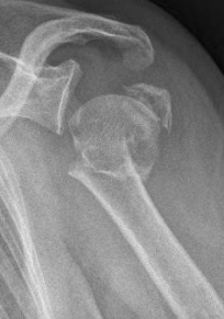
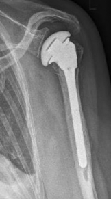
Picture: A subcapital upper arm fracture was treated with a prosthesis. The humeral head was completely removed and replaced by the prosthesis. This is anchored in the upper arm with the prosthesis shaft.
Fracture of the upper arm
Definition:
The upper arm shaft fracture is a fracture without involvement of the subcapital (shoulder joint) or supracondylar (elbow) region
Symptoms:
Malpositioning and often shortening in the upper arm area occur. Furthermore, a restraint of the arm is often to be found. The patient then supports, for example, the arm at the elbow with the other hand. Pressure and movement pain and a grinding of the bones are also typical. It is particularly important to rule out damage to the radial nerve and more rarely to the ulnar nerve in this type of fracture. The former is particularly endangered by its close proximity along the upper arm shaft. An injury is manifested by sensory disturbances on the forearm reaching the 1st - 3rd fingers on the extensor side.. Furthermore, vascular damage caused by palpation of the pulses at the elbow and wrist must be ruled out.
Diagnostics and Therapy:
In addition to the clinical symptoms already described, the cause of the accident must be ascertained. Either a direct impact of force has taken place, for example by a blow to the upper arm. A fall on the arm or, rarely, arm wrestling can be considered as an indirect force effect. This often leads to a so-called typical spiral fracture of the upper arm. X-rays of the upper arm including the adjacent joints must be taken in 2 planes.
The fractures of the upper arm are divided into simple, wedge and complex fractures according to the Workgroup for Osteosynthese. The above-mentioned frequent spiral fracture is one of the simple fractures, unless several fragments are present. Depending on the classification, usually upper arm fracture is treated.
Fractures which are not or only slightly displaced can be treated with a shoulder vest which immobilises the upper arm. The same applies to displaced simple fractures, which can be brought back into a correct position by pulling on the arm. This initial therapy can be supplemented or replaced by plaster treatment or splinting with a cast.
Severe shifted fractures, open fractures, multiple fractures and injuries to nerves and vessels are some of the most frequent indications for surgical therapy. Particularly in cases of suspected nerve injury, the quickest possible treatment must be sought in order to avoid consequential damage of nerve and movement disorders. Most frequently, the inner splinting of the upper arm is done by an intramedullary nail. Further possibilities are stabilization by means of plate osteosynthesis or, in the case of complicated fractures or many concomitant injuries, the attachment of an external fixator.
Follow-up treatment:
Upper arm fractures have a very good healing prognosis.
In conservative therapy, regular x-ray examinations are important to exclude secondary displacement of the fractures. To avoid this, the upper arm should be immobilised until complete bone healing is achieved. This is usually the case after 6 - 8 weeks. Afterwards, physiotherapeutic exercise is important to strengthen the muscles and restore mobility.
After surgical treatment, immediate exercise of the arm is possible. Here, too, physiotherapy and exercise is essential to strengthen the muscles in order to achieve a good surgical result.
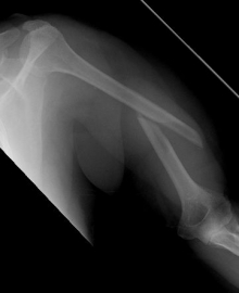
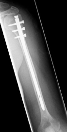
Picture: Here you can see a completely shifted upper arm fracture with a strong bend of the axis. First the operative treatment was performed by repositioning the bone and internal splinting of the fracture with a medullary nail.


Picture: An upper arm shaft fracture can alternatively be stabilized by a plate that bridges the fracture. The picture shows a shaft fracture with a bending wedge. After surgical treatment, the upper arm shaft is back in the correct axis.


